Have you found a concerning lump or bump on your horse? Skin tumors are common in all equine breeds and ages, but the specific lesion must be identified so the appropriate treatment is provided. Our Leatherstocking Veterinary Services team is therefore providing relevant information about four common equine dermatologic tumors and an explanation of how these lesions are treated.
Equine sarcoid
The most common tumor identified in horses is the equine sarcoid, which accounts for more than half of all equine skin tumors. These lesions are associated with bovine papillomavirus but need other factors for tumor growth. Other relevant information about equine sarcoids includes:
- Transmission — Sarcoids may be transmitted by contacting contaminated grooming equipment and tack, and may possibly be spread by flies.
- Location — These lesions can occur anywhere on the body, but the head, near the genitals, and scarred areas are the most common sites.
- Breed predilection — Quarter horses, Arabians, and Appaloosas are at increased risk for sarcoids.
- Types — Sarcoid types include:
- Occult — These lesions are flat, gray, hairless, and usually circular.
- Verrucose — Verrucose sarcoids appear scab- or wart-like and may contain small nodules.
- Nodular — These discrete, solid nodules may ulcerate and bleed.
- Fibroblastic — Fibroblastic sarcoids are irregular, fleshy, ulcerated growths that bleed easily.
- Mixed — Mixed sarcoids contain characteristics of two or more types.
- Malignant — These rapidly growing tumors spread extensively through the skin and infiltrate underlying tissue.
- Diagnosis — A presumptive diagnosis can often be made on clinical appearance alone. A definitive diagnosis requires histopathology, but biopsy-induced trauma can exacerbate the sarcoid.
- Treatment — Sarcoid treatment depends on the tumor type and location. Potential treatments include benign neglect for small occult or verrucous tumors, surgical excision, topical treatments to stimulate the immune system or target viral agents, chemotherapy, cryotherapy, immunotherapy, and radiation. In some cases, a multi-modal approach is used.
Equine squamous cell carcinoma
Equine squamous cell carcinoma (SCC) is the second most common skin tumor in horses. Exposure to chronic skin irritation and ultraviolet light contributes to tumor development, and equine caballus papillomavirus may also play a role. SCCs are usually locally invasive and slow to metastasize, although metastasis to local lymph nodes is not uncommon. Other relevant information includes:
- Location — Areas where SCCs occur include:
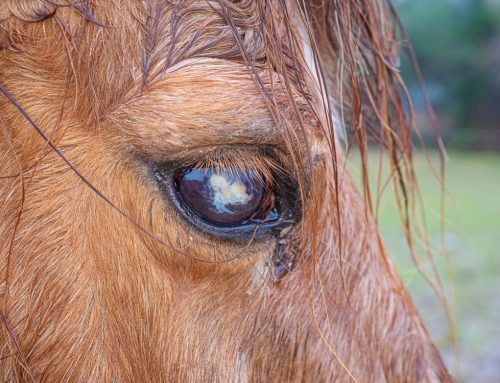
- Eye — The lesions can occur on the eyelids, third eyelid, outside corner, or on the corneal surface.
- Skin — Cutaneous SCCs can occur on the face, ears, and around the anus.
- Penis — Penile SCCs commonly affect older geldings. Stallions are rarely affected, which indicates that irritation from smegma may contribute to tumor formation.
- Vulva — Smegma may also contribute to vulva SCC formation in older mares.
- Mucosal — Mucosal surfaces lining the mouth and nose can be affected.
- Breed predilection — Paint horses, Quarter horses, Appaloosas, and draft horses are at increased risk for SCCs that surround the eye, and Quarter horses and Appaloosas are at increased risk for penile and vulvar SCCs.
- Diagnosis — Definitive diagnosis requires histopathology evaluation and possible local lymph node assessment for metastasis.
- Treatment — Treatment depends on tumor size and location and is most successful when initiated early or the lesion is small. Treatment typically involves surgical or cryosurgery removal, cytotoxic chemical application, or a combination of these methods. Surgical tumor removal with wide margins is the treatment of choice when location and size allow.
Equine melanoma
Melanomas are tumors that affect melanocytes, the skin cells responsible for producing skin and hair pigment. These tumors are extremely common in older grey horses, with more than 80% of grey horses having at least one melanoma during their lifetime. Melanomas that occur in non-grey horses are typically more malignant. Other relevant information about equine melanomas include:
- Location — Melanomas most commonly occur in the skin around the anus and the tail base. Other potential sites include:
- Eyelids, iris, and retina
- Mouth—especially the lips
- Parotid salivary glands and lymph nodes
- Penis and vulva
- Internal organs, including the intestine, heart, and lungs
- Breed predilection — Arabians, Lipizzaners, Adalusians, and Percherons seem more susceptible to melanomas, but this may be because they tend to be grey.
- Diagnosis — The melanoma’s clinical appearance is typically distinct enough for a presumptive diagnosis, but histopathology evaluation may be necessary for definitive diagnosis.
- Treatment — Small melanomas may not affect the horse, but lesions still should be monitored regularly, because they can grow quickly and cause significant problems. Potential treatments include surgical excision, cryosurgery, intralesional chemotherapy, and melanoma vaccines, which stimulate the horse’s immune response to the tumors.
Equine warts

Equine warts are a benign skin growth caused by the equine papillomavirus (EPV) that appear as single or multiple growths on the skin surface. They are most commonly seen in horses younger than 3 years of age. Warts are most commonly found on the muzzle and lips, but can also be found on the eyelids, ears, penis, vulva, mammary glands, and lower limbs. They typically resolve on their own in one to nine months and need no treatment. Quarantining the affected horse is recommended to prevent spreading EPV to other horses.
If your horse has a new lump or bump, contact our Leatherstocking Veterinary Services team, so we can investigate the lesion and determine an appropriate treatment plan.


Leave A Comment